| MEDICAL INTRO |
| BOOKS ON OLD MEDICAL TREATMENTS AND REMEDIES |
THE PRACTICAL |
ALCOHOL AND THE HUMAN BODY In fact alcohol was known to be a poison, and considered quite dangerous. Something modern medicine now agrees with. This was known circa 1907. A very impressive scientific book on the subject. |
DISEASES OF THE SKIN is a massive book on skin diseases from 1914. Don't be feint hearted though, it's loaded with photos that I found disturbing. |
3. TUBERCULOSIS VERRUCOSA
Verruca necrogenica, or anatomic tubercle, is the simplest and most
common of this rare form of cutaneous tuberculosis. It is a localized,
papillary or wart-like formation, occurring usually about the knuckles
or other parts of the hand or forearm. Its most common site is over one
of the metacarpophalangeal joints. It begins, as a rule, as a small,
papule-like growth, increasing gradually, but very slowly, in area, and
when well advanced, appears as a pea- or dime-sized or larger, somewhat
1 Colcott Fox, Brit. Jour. Derm., 1898, p. 253 (case demonstration).
2 Haushalter, Annales, 1898, p. 455.
3 Du Castel, ibid., 1898, p. 729.
4 Adamson, Brit. Jour. Derm., 1899, p. 20.
Among other pertinent interesting reports are: Bunch, “On Necrotic Tuberculides,”
Brit. Jour. Derm., 1912,p. 357 (with illustration), described 2 unusual cases: first, a boy
of twelve, in whom the disease began at the age of four as a simple red, slightly scaly
patch at navel on which a number of small nodules of a superficial papular character
appeared which gave place to shallow scars; there were also on the inner sides of thighs,
and around about the axillæ areas of a pinkish superficial dermatitis, which in the course
of several years became the seat of similar shallow scars, doubtless preceded by papula-
tion. The second case, a woman of twenty-five, presented a number of pea- and slightly
larger-sized pinkish indurated nodular swellings on the legs and arms; they gradually
broke down and showed slight purulent discharge; some coalescent in places, granulating
and healing, leaving considerable scarring; the disease began ten years previously; some
time after the appearance of these tuberculous lesions the patient developed a well-
marked lupus erythematosus.
Leopold and Rosenstern, “The Significance of Tuberculides in the Diagnosis of
Tuberculosis in Infancy,” Jour. Amer. Med. Assoc, Nov. 12, 1910, p. 1721, state that
from their experience in the Children’s Asylum in Berlin the small papulosquamous
and papulonecrotic tuberculides are not uncommon in cases of tuberculosis in infancy
(in 40 per cent, in their series); the lesions may be scanty and insignificant and unless
carefully searched for overlooked.
TUBERCULOSIS CUTIS
713
inflammatory, red, elevated, flattened, warty mass, with commonly a
tendency to slight pus-formation between the papillary projections, and
of which small drops can, as a rule, be made to ooze upon slight lateral
pressure. The surface, which is usually irregular, may be somewhat
hard or horny, or the growth may be covered with a crust. In fact,
the beginning papular lesion often becomes pustular, and this dries up
and forms a crusted covering, which, if removed by accident or design,
rapidly forms anew; later it acquires a warty aspect from papillary hy
pertrophy, and the same condition results as described above. The
border especially is often superficially indurated, rarely deeply. There
is also a disposition toward central healing and peripheral spread,
although the patch rarely reaches more than an inch or so in area. The
growth is more or less persistent, but in some cases, after a variable time,
undergoes involution, leaving usually a slight smooth or corded scar.
While the lesion is, generally speaking, relatively benign, it is occasion
ally followed by extension along the lymph-channels, with involvement
of the lymphatic glands and constitutional infection; according to Knick-
enberg,1 Guizzetti,2 Hallopeau,3 and others, this occurs more readily in
this form than in that known as tuberculosis verrucosa cutis (of Riehl
and Paltauf). Its usual subjects are those who have to do with decom
posing animal matter; hence it is chiefly encountered among medical
students, physicians, dissection-room and hospital-ward attendants, and
butchers. Besnier and Vidal originally noted its occurrence among those
making autopsies in tuberculous subjects, and recognized its analogy to
some types of lupus. The investigations of Riehl,4 Finger,5 and others
demonstrated that verruca necrogenica is due to tubercle bacilli infection.
Tuberculosis verrucosa cutis (of Riehl and Paltauf), while for some
years irregularly met with, was first brought into prominent notice
by the classic paper by Riehl and Paltauf,6 whose studies were based
upon 14 cases observed in Kaposi’s clinic, and which showed the analogy
and probable identity of this with anatomic tubercle just described;
also its resemblance to lupus verrucosus. It was by them thought to
occur in adults who had to do with animals and animal products, but we
know today that many cases are also contracted from other tuberculous
individuals. White7 believes that this variety may fairly be accepted
as the ordinary form of manifestation in the cutaneous tissues of the
inoculation of the same with tuberculous matter, whether derived from
the cadaver of man or other animal, or by contact with infective material
from living subjects. While it usually begins in an insignificant manner,
practically similarly to verruca necrogenica, it is much more extensive
and may cover several inches or more of surface. One, several, or more
1 Knickenberg, “Ueber Tuberculosis verrucosa cutis,” Archiv, 1894, p. 405.
2 Guizzetti, “Ueber einen Fall von Tuberculum anatomicum. Histologisch-bak-
teriologische Untersuchungen,” Monatshefte, 1899, vol. xxix, p. 253.
3 Hallopeau, “Sur les rapports de la tuberculose avec les maladies de la peau autres
que le lupus vulgaris,” Jour. mal. cutan., 1896, p. 522.
4 Riehl and Paltauf, “Tuberculosa verrucosa cutis,” Archiv, 1886, p. 19.
5 Finger, “Ueber die sogenannte Leichenwarze,” Deutsche med. Wochenschr., 1888,
p. 85. 6 Loc. cit.
7J. C. White, “Clinical Aspects and Etiologic Relations of Cutaneous Tubercu
losis,” Boston Med. and Surg. Jour., Nov. 12, 1891, p. 509.
714
NEW GROWTHS
small patches arise, and if the latter, generally near together, and sooner
or later coalesce and form an irregularly shaped or serpiginous area.
The degree of inflammatory action present varies considerably, the area
or areas being surrounded by a band-like redness with some infiltration.
The whole patch is, as a rule, somewhat elevated, infiltrated, and beset
with wart-like papillary hypertrophy, or with dull or purplish-red tuber
cles or nodules, isolated or grouped; or with closely aggregated or discrete,
usually minute pustules, seated directly upon the sluggishly inflamed

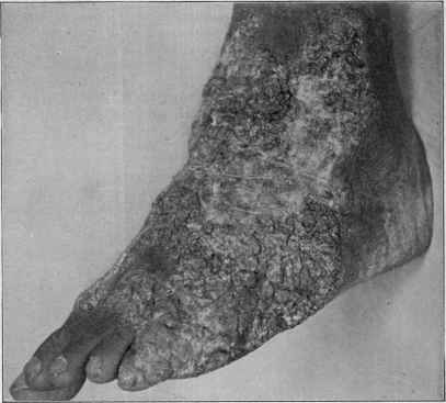
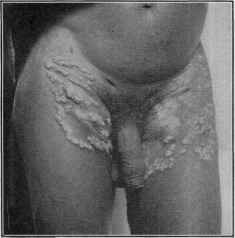
Fig. 158.—Tuberculosis verrucosa cutis; in a negro.
purplish area, or at the summits of the tubercles or nodules. Not in
frequently the interstices between the papillary or projecting vegetations
contain some seropurulent or purulent fluid which can be pressed out.
While the disease spreads peripherally, a slight tendency to thin scarring
is generally to be noted in the earliest portions. In fact, in some cases,
especially where there are several areas or one extensive plaque, all the
described features can usually be seen. One of its characteristics is
that it rarely shows any positive ulceration.
Its favorite region is the back of the hand—sometimes one, sometimes
both. It often spreads on to the fingers and the interdigital folds, up
the wrists, and less frequently on to the palms. On this last-named
region it is, however, rare, and always appearing as an extension from
TUBERCULOSIS CUTIS
715
the dorsum, as in the 2 cases reported respectively by Cutler1 and Weber.2
Inasmuch as this type is frequently seen in association with pulmonary
phthisis, 4 examples of which have been recently reported by Béclère,3
it is probable, as stated by this observer, following Vidal, that its presence
in this region finds its explanation in the fact that phthisical patients,
and especially men, in whom this manifestation is most frequently en
countered, after coughing wipe off the mouth and mustache with the
back of the hand. Of interest and suggestive in this connection is
Schoull's4 experiment, who took some hair from the beard of a tuberculous
patient, and, after soaking and agitating it in distilled water in a test-
tube, injected some of the liquid into a guinea-pig, the animal shortly
succumbing to tuberculosis. Fabry5 called attention to its occurrence
in coal-miners, in whom the numerous and constant injuries and cuts
about the backs of the hand incident to their work play an important

Fig. 159.—Tuberculosis verrucosa cutis. Patient aged twenty-four years; duration four
years; sinuses of knee (courtesy of Dr. F. C. Knowles).
rôle, affording opportunities for inoculation, phthisis being not an un
common, disease among such workmen.
Like anatomic tubercle, it is slow in its course, often stationary for
periods, and in certain parts retrogressive; it often lasts for years, and
occasionally disappears spontaneously. There is usually left a thin,
atrophic-looking scar, sometimes with the sieve-like aspect noted in
lupus erythematosus. It bears a striking resemblance to blastomycetic
1 Cutler, Jour. Cutan. Dis., 1898, p. 535 (case demonstration)—on backs and palms
of both hands.
2 Weber, Brit. Jour. Derm., 1899, p. 161 (case demonstration).
3 Béclère, Gaz. hebdom., 1898, No. 34; Annales, 1898, p. 794.
4 Schoull, Jour, des prat., 1899, p. 347.
5 Fabry, “Ueber das Vorkommen der Tuberculosis verrucosa cutis (Riehl and
Paltauf) bei Arbeitern in Kohlenbergwerken,” Archiv, 1900, vol. li, p. 69 (7 cases, most
of which with associated pulmonary tuberculosis, or with the latter in the family; 2
cuts of cases); and Schulze (Fabry’s clinic), ibid., 1904, vol. lxx, p. 329 (a synopsis of
160 cases).
7l6 NEW GROWTHS
dermatitis (q. v.), with which doubtless it has sometimes been con
founded.
Various other forms allied to this in general character have been
described, but they are scarcely to be considered as separate manifesta
tions, but rather as aberrant types, in which one or other clinical element
predominates, and sometimes presenting features partaking of the nature
of both this form and lupus vulgaris; moreover, it may occasionally be
upon other situations. Thus, in Morrow’s1 case (tuberculosis papillo-
matosa cutis) the eruption was seated upon the face, and consisted of
rather bright-red, abundant, and extremely prominent papillomatous
vegetations; in Mracek’s2 and Ravogli’s3 cases, the leg was the seat of
the manifestation, which was quite extensive.
In some rare cases, described by Riehl4 as tuberculosis fungosa
cutis, the growths are somewhat fungoidal or mushroom-like, and re-

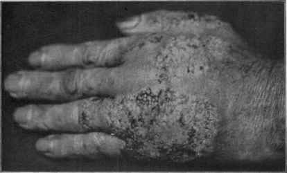
Fig. 160.—Tuberculosis verrucosa cutis in patient with pulmonary tuberculosis. (Note
the close resemblance to blastomycosis.)
semble similar formations observed in granuloma fungoides; the disease
usually originates in osseous or periosteal lesions; fistulous ulcers are
formed, growing nodular swellings, with fungoidal aspect, and later the
ordinary appearances of tuberculous ulcers. Other lesions, occasionally
seen in the average type, are tubercles or nodules, small pustules, papil
lary elevations, and crusting. In other instances, as described (fibroma-
tosis tuberculosa cutis) by the same author, along with papillomatous
vegetations, ulcers, etc, there is marked sclerosis found on different
1 Morrow, Jour. Cutan. Dis., 1888, pp. 361 and 401 (with colored plate); Shelmire,
Jour. Cutan. Dis., 1906, p. 20, reports a case of probably a mixed type, the verrucose
element predominating; eruption on face and neck in a negress (2 good illustrations).
2 Mracek, Hand Atlas of Skin Diseases, second edit., plate 86.
3 Ravogli, “On Various Forms of Cutaneous Tuberculosis,” Jour. Amer. Med.
Assoc., April 16, 1898.
4 Riehl, Verhandl. der Deutschen dermatolog. Gesell., iv Congress, 1894, p. 354.
TUBERCULOSIS CUTIS
717
parts, not infrequently about the nose, mouth, and other mucous outlets.
Wickham and Jessner have also described cases with frambesiform
lesions, in that of the latter the eruption being disseminated. In fact,
almost all types of nodular, vegetating, and ulcerative lesions are en
countered as the result of tubercle bacilli infection, and Besnier, Lejars,
Jeanselme, and others have noted rare instances (lymphangitis tubercu-
losa cutanea)1 in which the lymphatics, usually of the extremities, were
the seat primarily or secondarily of infection, and the eruptions of various
characters, lupoid, nodular, and papillomatous, corresponding to the
lymphatic distribution, and commonly with some elephantiasic enlarge
ment. To lymphatic involvement is doubtless due also the hypertrophic
character noted in other types.
Diagnosis.—The sluggish character of the growth, its dull red
or purplish-red color, its localization, history, and slow course, and the
papillomatous tendency displayed, the absence of distinct ulcerative
action, and usually of brownish or reddish-yellow tubercles, will serve
to distinguish it from both syphilis and other forms of tuberculosis.
Its resemblance to blastomycetic dermatitis is striking, and, in fact,
unless other symptoms of tuberculous nature are present, a conclusive
differentiation is possible only by microscopic and bacteriologic means.
Treatment.—Of the various local measures to be mentioned in
the section on treatment of the tuberculoses, the use of the antiseptic
applications, such as the mercurial preparations, together with the use
of strong salicylic acid plasters and pyrogallol salves, and, when neces
sary, the curet, are the most valuable. The x-ray and Finsen treat
ments have also been found serviceable.
But first, if you want to come back to this web site again, just add it to your bookmarks or favorites now! Then you'll find it easy!
Also, please consider sharing our helpful website with your online friends.
BELOW ARE OUR OTHER HEALTH WEB SITES: |
Copyright © 2000-present Donald Urquhart. All Rights Reserved. All universal rights reserved. Designated trademarks and brands are the property of their respective owners. Use of this Web site constitutes acceptance of our legal disclaimer. | Contact Us | Privacy Policy | About Us |