| MEDICAL INTRO |
| BOOKS ON OLD MEDICAL TREATMENTS AND REMEDIES |
THE PRACTICAL |
ALCOHOL AND THE HUMAN BODY In fact alcohol was known to be a poison, and considered quite dangerous. Something modern medicine now agrees with. This was known circa 1907. A very impressive scientific book on the subject. |
DISEASES OF THE SKIN is a massive book on skin diseases from 1914. Don't be feint hearted though, it's loaded with photos that I found disturbing. |
Pigmentary Syphiloderm1 (Synonyms: Syphiloderma pigmento-
sum; Syphilitic leukoderma; Vitiligo acquisita syphilitica)—This is a rare
manifestation about the correct nosology of which there has been much
difference of opinion. It is now pretty generally conceded, however,
that it is of syphilitic origin, although some authors still maintain that
it has no direct relationship to this disease. While first described by
Hardy in 1853, it was not until Fournier’s presentation of it (1873)
that it received much attention. Since then various observers, among
whom G. H. Fox, Atkinson, Taylor, Maireau, Pœlchen, Malherbe,
Neisser, and Maieff have reported cases or contributed special papers.
It is essentially a macular eruption, although totally unlike the macular
syphiloderm as commonly met with and just described; the former is one
of pigmentary changes pure and simple, the latter due to hyperemia.
It appears during the earlier secondary stage or toward the end of the
first year, although it sometimes does not present until a later period.
The region of the neck and shoulders is its usual location, Fournier stating
1 Principal literature: Hardy, Maladies de la peau, Paris; Fournier, Lecons sur la
syphilis, étudiée plus particuliérment chez la femme, Paris, 1873, P- 422 (with colored
plate); G. H. Fox, “On the So-called Pigmentary Syphilid,” Amer. Jour. Med. Sci.,
April, 1878; Atkinson, “The Pigmentary Syphiloderm,” Chicago Med. Jour, and Exam.,
1878, vol. lxxxvii, p. 340; Neisser, “Ueber das Leucoderma syphiliticum,” Archiv,
1883, p. 491; Taylor, “On the Pigmentary Syphilid,“ Jour. Cutan. Dis., 1885, p. 97;
Pœlchen, “Vitiligo acquisita syphilitica,” Virchow’s Archiv, 1887, vol. cvii, p. 535
(with 2 colored plates); Malherbe, “Deux cas de syphilide pigmentaire chez l'homme,”
Gazette méd de Nantes, Dec 12, 1895, p. 13 (2 cases), abs. in Annales, 1896, p. 968;
Maieff, “Contribution a l’étudè de la syphilide pigmentaire,” Trans. 1nternat. Dermat.
Congress, Paris, 1889, p. 677 (with bibliography); Maireau, “Syphilide pigmentaire,"
These de Paris, 1884 (with literature references); Lang, Vorlesungen über Pathologie und
Therapie der Syphilis, Wiesbaden, 1896, p. 208 (with cut); Ehrmann, “Ueber Haut-
färbung durch secondar-syphilit. Exanthemata,” Archiv, 1891, p. 79.
syphilis 779
that in but 1 in 30 cases is it found elsewhere than on the neck, although
it may also, however, exceptionally invade other parts.
According to Taylor, three forms are encountered: (1) as spots or
variously sized brownish patches; (2) more or less diffused brownish
discoloration, which subsequently becomes the seat of small, spotty,
leukodermic changes, which increase in size, and the general appearance
of which is retiform (retiform pigmentary syphiloderm or syphilid);
(3) an abnormal or uneven distribution of pigment, the surface having
a dappled or marmoraceous aspect (marmoraceous pigmentary syphilid).
The first, spot or patchy form, varies in color between a light and dark
brown, and the spots or patches are rounded or oval, sometimes with
irregularly jagged edges, and not commonly with uniform pigmentation,
the bordering part frequently showing the deeper shade. Intervening
white skin looks relatively of diminished normal pigmentation. The
second or diffused form is the most usual type encountered, beginning at

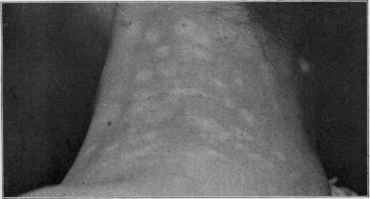
Fig. 176.—Pigmentary syphiloderm (neck and shoulders); was first diffused pig-
mented, the vitiligo-like spots subsequently appearing (syphilitic leukoderma); pre
sented about the sixth to eighth month of the disease. Patient a woman.
the neck, especially at the sides, where it may remain, or it may invade
the trunk and arms. Its appearance may be rapid or gradual. Sooner
or later white points or spots show themselves, and the condition is some
what suggestive of leukoderma. Generally it is this change which first
calls the attention of the patient to the existing discoloration. The
third variety is the rarest of all, and its advent is insidious, and is always
seen (Taylor) on the sides of the neck, with no tendency to spread.
According to Taylor, there is no hyperpigmentation, primarily at least,
but the process is more that of irregular pigment absorption, the inter
vening remaining normally pigmented spots appearing dark by compari
son; other observers, however, have noted the contrary. For some time
the manifestation was considered to occur in women only, but this is
now known to be erroneous, as it has been also observed, although much
less frequently, in males by Chambard,1 Malherbe (loc. cit.), and others.
It is much more common in brunettes.
1 Chambard, cited by Crocker, Diseases of the Skin,
780
NEW GROWTHS
There is a diversity of views as to whether this eruption, if it may
be so called, arises as such or is in reality a vitiligo of syphilitic origin,
originating in the spots of a preceding syphiloderm (Fox, Lang, Neisser,
Pœlchen). Its duration is variable,—from a few months to several years
or more,—it is without subjective symptoms, and is wholly uninfluenced
by antisyphilitic remedies, in this respect differing from all other syphilitic
manifestations; and this last fact, it must be confessed, gives some
grounds for at least questioning whether it is a syphilitic manifestation
sui generis, or a chloasmic condition dependent upon a syphilitic cachexia
or upon a previous evanescent, ordinary, macular syphiloderm. In
some instances of syphilodermata, usually in the late secondary stage,
may be seen dark blue or livid spots on the trunk chiefly, interspersed
among the eruptive lesions; Ehrmann (1907), who first described them
and gave the name Livedo racemosa, thought them due to endothelial
proliferation in the arterioles, interfering with the blood-current.1
In the diagnosis care is to be exercised that the pigmentary syphilo-
derm be not confused with ordinary chloasma, vitiligo, and tinea versi-
color. Its usual limitation to the neck, with little if any tendency to
appear in other situations, is unusual with these several affections;
tinea versicolor, moreover, always involves at least the upper chest region
as well, and its slight furfuraceous or branny scaliness noted when the
skin is dry is another differential factor to which many more would, if
necessary, readily suggest themselves by referring to the description of
that malady.
But first, if you want to come back to this web site again, just add it to your bookmarks or favorites now! Then you'll find it easy!
Also, please consider sharing our helpful website with your online friends.
BELOW ARE OUR OTHER HEALTH WEB SITES: |
Copyright © 2000-present Donald Urquhart. All Rights Reserved. All universal rights reserved. Designated trademarks and brands are the property of their respective owners. Use of this Web site constitutes acceptance of our legal disclaimer. | Contact Us | Privacy Policy | About Us |