| MEDICAL INTRO |
| BOOKS ON OLD MEDICAL TREATMENTS AND REMEDIES |
THE PRACTICAL |
ALCOHOL AND THE HUMAN BODY In fact alcohol was known to be a poison, and considered quite dangerous. Something modern medicine now agrees with. This was known circa 1907. A very impressive scientific book on the subject. |
DISEASES OF THE SKIN is a massive book on skin diseases from 1914. Don't be feint hearted though, it's loaded with photos that I found disturbing. |
ACTINOMYCOSIS
Synonyms.—Actinomycosis of the skin; Lumpy jaw; Fr., Actinomycose; Ger.,
Aktinomykose.
Definition.—Actinomycosis of the skin is an affection due to
the ray fungus, characterized by a sluggish, red, nodular, or lumpy in
filtration, usually with a tendency to break down and form sinuses, and
most commonly involving the cervicofacial region.

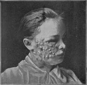
Fig. 305.—Actinomycosis (courtesy of Dr. W. T. Corlett).
The condition known as lumpy jaw and osteosarcoma of the jaw
in cattle had long been known, but it was Israel who first recognized the
pathogenic rôle of the special fungus, named by Harz the ray fungus.
About the same time the existence of a similar looking affection in man
was described by Israel, and which was subsequently shown by the im
portant contribution by Ponfick to be not only similar to that in animals,
but of identical nature. Since then the malady and its fungus have
received considerable attention from various observers, among whom are
Illich, Majocchi, Bertha, Gasperini, Krause, Müller, Poncet and Bérard,
1156
PARASITIC AFFECTIONS
Murphy, and many others.1 While the fungus may gain access to the
internal organs and give rise to grave disease, the dermatologist is chiefly
interested in the manifestations observed when the integumentary
tissues are invaded. The invasion of the latter may be primary, but, as a
rule, it is secondary to a deeper-seated involvement.
Symptoms.—The usual situation of actinomycosis of the skin is
about the jaw, neck, and face. The organism finds entrance through
the mouth, most frequently to the jaw through a decayed tooth. The
first evidence is a hard, subcutaneous swelling or infiltration, which
may attain moderate or quite conspicuous dimensions, the overlying
skin soon becoming of a sluggish or dark-red color. Sooner or later
softening is detected, the skin giving way at one or several points, from
which there oozes a discharge of a seropurulent, purulent, or sanguino-
lent and purulent character. Contained in the discharge, recognizable in
most instances, are minute, friable, yellowish or yellowish-gray bodies,
representing conglomerate collections of the fungus. Instead of begin
ning or continuing as a well-defined single swelling or tumor, the in
volved and infiltrated area is distinctly nodular, often finally becoming,
when at all advanced, quite extensive. It is then noted to consist of a
variously and irregularly infiltrated and swollen area, dark red or bluish
red, beset with several or more distinct nodulations or anthracoid for
mations, with here and there openings leading down or through the in
volved mass, with slight, moderate, or profuse discharge.2 In some cases
the surface exhibits ulcerofungoidal and papillomatous characters.
Occurring on other parts of the body the same conditions are presented,
occasionally involving considerable surface.3 Sometimes it remains
limited to a more or less circumscribed area,4 several finger cases
having been recorded.
The course of the malady may be slow and insidious, or somewhat
rapid, usually the former, some months generally elapsing before the
involvement is extensive. As a rule, there are no subjective symptoms,
but when suppuration takes place the parts may become quite painful.
The lymphatic glands are not implicated except secondarily as a result
of the suppurative inflammation. The general health in those instances
1 Poncet and Bérard’s monograph, Traité clinique de V actinomycose humaine, Paris,
1898, gives an admirable and exhaustive presentation and review of the subject, with
complete bibliography.
2 Wallhauser, Jour. Cutan. Dis., 1904, p. 77 (with illustration), reports an extensive
case of this kind beginning as a small pimple on point of chin, and gradually involving
the whole regions of the upper part of the neck and the jaws.
3 Pringle, London Med.-Chir. Soc’y Trans., 1895, vol. lxxviii, p. 21 (with colored
case illustration), reports an extensive case in a boy of eleven, implicating part of the
chest, the back, and hip, and developing secondarily to involvement of the pleura.
4 Sicard, La presse médicale. Aug. 15, 1903, reports a case in which it was confined to
the finger, and the earliest symptoms (following an accidental cut in a field-worker)
were of a vesicular character; Massaglio, ibid., Aug. 31, also a finger case; Thevenot,
ibid., 1903, vol. 1xxvii, p. 659, reports a case of a nodular type of paronychia of the finger
caused by the actinomyces; Wright, Amer. Jour. Med. Sci., July, 1904, p. 74, a tonsil
case.
Some later general papers: Sawyer, Jour. Amer. Med. Assoc, March 11, 1901;
Ewing, Bull. Johns Hopkins Hospital, Nov., 1902; von Baracz, Annals of Surgery,
March, 1903—abs. in Jour. Amer. Med. Assoc, March 21, 1905: Howard, Jour. Med.
Research, 1903, vol. ix, p. 301; Dor (researches on fungus), La presse mêdicale, Sept. 16,
1903; Stokes, Amer. Jour. Med. Sci., Nov., 1904, p. 861; Knox, Lancet, Oct. 29, 1904.
A CTINOM YCOSIS 1157
where the invasion is from a superficial part ordinarily remains unin
fluenced unless systemic pyemic infection occurs or the fungus elements
find their way to the deeper organs or structures.
Etiology and Pathology.—The disease is due to the ray fungus.
It is somewhat rare, and apparently oftener observed in Germany and
France than elsewhere. The first cases described in our own country
are those by Murphy (1885), Schirmer, Ochsner, and Bodamer (1889),1
It is contagious by inoculation, and commonly contracted from cattle
and horses, and therefore seen most frequently in those who have to do
with these animals. It is probable, too, that, in some instances, as in
that noted by Baracz2 from kissing, it may be communicated from one
individual to another. As the fungus is also believed to flourish on
straw, corn, and other grain, the habit among farmers, dairymen, and
others of chewing upon such substances3 is very likely responsible for the
common method of inoculation through the mouth, taking place, as a
rule, through a decayed tooth. According to Lord,4 actinomycetes can
be demonstrated in the contents of carious teeth and the crypts of the
tonsils in persons without actinomycosis, indicating that the buccal cavity
may be a possible source of the disease. The fungus has also been found
in bovine vaccine virus (Howard).5 Successful inoculation ordinarily
presupposes an abrasion or break of continuity, and this has usually
been noted in those instances, relatively few, in which the integument
was primarily involved.6 In most of these latter cases the area involve
ment was small.
1 Bodamer’s paper, Med. News, March 2, 1889, gives abstract of the others, with
references.
2 Baracz, Wiener med. Presse, 1889, p. 6 (man to wife).
3 Ljunggren, Nordiskt med. Arkiv, 1895, No. 27, P. 1—brief abs. in Annales, 1896,
p. 763, refers to 27 cases (13 personal) occurring in those in the habit of chewing grain
or straw; Zeisler’s case. Jour. Cutan. Dis., 1906, p. 510, was attributed to the chewing
of grass; Varney’s case, ibid., 1909, p. 235 (systemic, neck, cheek, and leg; ray fungus
found in the sputum), had been in the habit of chewing wheat kernels whenever he
could obtain them.
4 Lord, “A Contribution to the Etiology of Actinomycosis: Experimental Produc
tion of Actinomycosis in Guinea-pigs Inoculated with the Contents of Carious Teeth,”
Boston Med. and Surg. Jour., July 21, 1910; and “The Etiology of Actinomycosis;
The Presence of Actinomycetes in the Contents of Carious Teeth and the Tonsillar
Crypts of Patients Without Actinomycosis,” Jour. Amer. Med. Assoc, Oct. 8, 1910,
p. 1261.
5 Kendall (Australasian Med. Gaz., Feb. 1, 1913, p. 108; review editorial), at a re
cent Congress in Melbourne, stated that during the last few years over 600 cases of
actinomycosis of the udder had been met with in the dairy herds of Victoria and that
actinomycosis of other parts was also common.
6Kopfstein, Wiener klin. Rundschau, 1901, p. 21, reports the case of a woman, a
farm laborer, who developed the disease in the hand, presumably inoculated while bind
ing corn, through a cut accidentally made a few days previously. He refers also to
Müller’s case, in which infection was apparently due to the entry of a splinter of wood
into the palm of the hand; and another instance (Von Partsch) where it followed a sur
gical operation, inoculation occurring apparently by means of the surgeon’s instruments.
Merian, “Ein Fall von primarer Hautaktinomykose,” Dermatolog. Wochenschr., 1912,
vol. 1vi., p. 45, reports a case, nineteen-year-old girl, of primary skin infection occurring
in the left nasolabial fold at its lower part; the lesion being pea-sized, with a reddish-
blue zone; the growth was soft, and with slight yellowish-red pus oozing from its apex;
began, according to the patient, three weeks previously as a red itching spot about the
size of a hemp-seed. Several important papers on the disease are mentioned, with
references. This case, according to the author, makes about 25 cases of primary skin
infection to be found in literature; brief review with references.
1158
PARASITIC AFFECTIONS
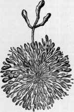
The fungus, called the actinomyces, consists of a central network
mass of interwoven threads, from which threads, or mycelia, radiate
like projecting rays, and terminate in bulbous expansions; these latter
are thought to represent the fructifying bodies. One to several may pro
ject beyond the others. In tissue-section examination sometimes small,
oval, apparently homogeneous bodies are seen lying near the ray fungus,
and suggestive of spore forms of the
organism (Rosenberger),1 While pre
viously the fungus was thought to be
long to the molds, Bostroem’s (1885)
investigations seemed to show it to be a
variety of cladothrix, of the class schizo-
mycetes, although on this point there is
still uncertainty and difference of opinion.2
Of the various staining methods for its
demonstration, that of Gram seems to be
most generally satisfactory. The fungus
is usually readily demonstrable, both in
the discharge (the yellowish grains) and
in sections of involved tissue. In some
instances, however, especially in the
earlier stages, it is not always found
(Legrain, Mackenzie, Knox, Galloway,
and others),3 and exceptionally only in
the sections from the outlying invading
borders.4
Histologically, the nodular and infiltrated mass is made up of granu
lation tissue having a resemblance to that of round-celled sarcoma; in
some instances epithelioid giant-cells and mast-cells are to be seen.
Diagnosis.—The disease is to be distinguished from syphilis,

Fig. 306.—Actinomyces, show
ing the ray arrangement and the
club-shaped ends of the mycelial
threads (after Ponfick).
1 Rosenberger, Jour. Applied Microscopy, Nov., 1900, vol. iii, p. 1051.
2 In an interesting paper on the biology of the micro-organisms, J. H. Wright, publi
cation of the Mass. Gen’l. Hosp., 1905, vol. i, No. 1, thinks from his studies and re
view that the widely disseminated branching micro-organisms thought by Bostroem
and others to be the specific infectious agent of actinomycosis are really quite different,
having spore-like reproductive elements, and should be grouped together as a separate
genus with the name Nocardia and that infection by them should be called nocardiosis,
and not actinomycosis; that the term “actinomycosis” should be used only for those
inflammatory processes the lesions of which contain the characteristic granules or
“drusen,” composed of dense aggregates of branched filamentous micro-organisms and
of their transformation or degeneration products—these products including the charac
teristic refringent club-shaped bodies radially disposed at the periphery of the granule,
and which may or may not be present. Apropos of this may be mentioned a recent
paper by Kieseritzky and Gerhardt, Archiv. klin.Chirurg., 1905, pp. 835, et seq., which
shows that some cases clinically resembling the disease, and even containing radiating
filaments are rather negatived by more careful, especially laboratory, investigation;
Pernet also shows (Brit. Jour. Derm., 1905, p. 265), in some cases clinically present
ing the picture of actinomycosis, the microscopic examination discloses the character
istic appearances of streptothrix.
3 Legrain, Annales, 1891, p. 772; Mackenzie, Brit. Jour. Derm., 1894, p. 370; Knox,
Glasgow Med. Jour., 1896, vol. xlv, p. 382; Galloway, Brit. Jour. Derm., 1895, p. 116.
4 In a case of a physician, involving the arm, operated upon several times at the
Jefferson Medical College Hospital, repeated examinations of the discharge and tissue
from the main portion failed to disclose the fungus, but it was finally found in sections
from the extreme outer edge of the spreading border.
ACTINOMYCOSIS
1159
sarcoma, carcinomata, tuberculous affections, mycetoma, and phleg-
monous inflammation. The presence of the peculiar yellowish bodies or
granules in the discharge would be of conclusive import. The common
location about the angle of the lower jaw and neck and cheek, and espe
cially occurring in those who have to do with animals and grain products,
should always lead to the suspicion of actinomycosis, which can be
verified or disproved by observation and by examinations for the fungus;
in doubtful instances repeated examinations should be made for the
latter in the discharge, and also in the deeper bordering tissue, before its
absence can be accepted as proved.
Prognosis.—Actinomycosis of the skin and superficial parts is
usually a remediable disease, although always fraught with the possi
bility of deeper involvement and grave consequences. Schlange,1 from
a study of. a number of patients, takes a rather favorable view of these
cases, stating, from his analytic study, that, excepting when involving
the internal organs, it has a pronounced tendency to spontaneous recov
ery. It does, however, in some instances continue almost indefinitely
without exhibiting such disposition. The advent of pyemic symptoms
is always of serious, and usually fatal, import. Involvement of the
upper jaw is more serious than that of the lower jaw or other surface
situations, as there is more danger of deep invasion.2 Involvement of the
orbit is also of serious portent. It is a matter of observation that some
cases are inherently mild and others more or less malignant, doubtless
due to the virulence of the fungus and the varying resisting power of
different individuals, and on the influence of accidental secondary in
fective processes.
Treatment.—The management of this malady consists in the
administration of potassium iodid in moderate or large dosage, con
jointly with, in obstinate or spreading cases, curetting or excision of the
diseased mass. This remedy varies in its effect in different instances,
but it has proved beneficial or curative (Carless, Pringle, Morris, Rydy-
gier, Jurinka, Nocard, Netter, Dubreuilh, Audry, Ljunggren, Claisse,
Bérard, and many others) in many cases, and should always be given a
good trial before operative measures are instituted. According to Bérard
and others, its most rapid and brilliant results are in those instances
in which the malady is recent and uncomplicated, but when there is
associated secondary infection by streptococci, staphylococci, or the
bacterium coli commune the remedy is less satisfactory. Rydygier3
successfully treated 2 cases by local injections of a 1 per cent, solution of
potassium iodid and sodium iodid, injecting at first one Pravaz syringeful,
later half as much again; one of these cases had been previously treated
without result by surgical means and the internal administration of the
drug. Bevan4 and Zeisler report favorably of copper sulphate, ¼-grain
(0.017) doses four times daily.
1 Schlange, “Zur Prognose der Aktinomykose,” Verhandl. f. Deutsch. Gesellsch. f.
Chirurg., 1892, part ii, p. 24.
2 See paper by Bourquin and de Quervain, “Sur les complications cérébrales de
l’actinomycosis,” Rev. méd, de la suisse rom., 1897, vol. xvii, p. 145 (with references).
3 Rydygier, Wien. klin. Wochenschr., 1895, p. 649; also Sawyer, loc. cit.
4 Bevan, Jour. Amer. Med. Assoc, Nov. 11, 1905.
1160
PARASITIC AFFECTIONS
The local treatment of the lesions is essentially that of similar nodular
ulcerative and suppurative formations—the maintenance of cleanliness
and the applications of mild antiseptics, a frequently changed wet dress
ing of LugoPs solution being one of the best, and probably of some
direct inhibitory influence upon the growth or effects of the fungus.
I have found the x-ray valuable.
But first, if you want to come back to this web site again, just add it to your bookmarks or favorites now! Then you'll find it easy!
Also, please consider sharing our helpful website with your online friends.
BELOW ARE OUR OTHER HEALTH WEB SITES: |
Copyright © 2000-present Donald Urquhart. All Rights Reserved. All universal rights reserved. Designated trademarks and brands are the property of their respective owners. Use of this Web site constitutes acceptance of our legal disclaimer. | Contact Us | Privacy Policy | About Us |