| MEDICAL INTRO |
| BOOKS ON OLD MEDICAL TREATMENTS AND REMEDIES |
THE PRACTICAL |
ALCOHOL AND THE HUMAN BODY In fact alcohol was known to be a poison, and considered quite dangerous. Something modern medicine now agrees with. This was known circa 1907. A very impressive scientific book on the subject. |
DISEASES OF THE SKIN is a massive book on skin diseases from 1914. Don't be feint hearted though, it's loaded with photos that I found disturbing. |
URTICARIA PIGMENTOSA1
Synonyms.—Xanthelasmoidea; Urticaria perstans pigmentosa.
Definition.—An urticaria-like eruption, in which the lesions are
usually persistent and show accompanying or subsequent pigment
deposit, with, in some, a new growth element.
1 Literature: For cases, etc, antedating 1883 (Nettleship, Morrant Baker, Tilbury
Fox, Barlow, Sangster, Morrow, Goodhart, Mackenzie, Cavafy) see paper by Colcott
Fox, Trans. Med.-Chir. Soc'y, London, 1883, p. 329; also Crocker‘s paper, Trans.
Clin. Soc'y, London, 1885, p. 12; also Raymond, “Urticaire pigmentée,” These de
Paris, 1888 (giving a complete review of the subject—referring to 29 cases). Among the
many cases reported since may be mentioned: Elliot, Jour. Cutan. Dis., 1891, p. 296;
URTICARIA PIGMENTOSA
189
Symptoms.—The eruption, which, with rare exceptions, makes its
appearance in the first several months of life, is, as a rule, scarcely
distinguishable in its beginning from ordinary urticaria; but the lesions,

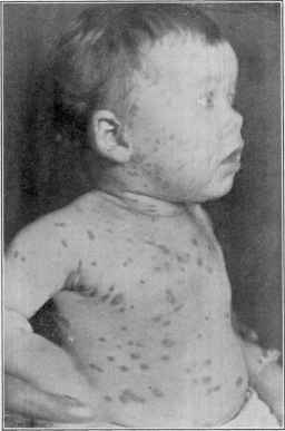
Fig. 36.—Urticaria pigmentosa in a female child eleven months old. Began in the
fourth month, at first on the legs, and gradually invaded the entire surface, including the
scalp. The lesions present as ordinary wheals, but are persistent, subsiding somewhat,
becoming yellowish, later with a violaceous tinge; after continuing for several months
or longer they disappear, leaving purplish and bluish-yellow stains. Some of the lesions
have a slight resemblance to xanthoma. Itching was, as a rule, moderate, only occa
sionally troublesome. Several years later the malady was much less active, and the
lesions relatively sparse. General health excellent.
or the most of them, instead of disappearing quickly, are persistent,
and after some days or a few weeks show pigment deposit. An individual
Hallopeau (leaving white cicatrices), Annales, 1892, p. 628; Bronson (soc‘y discus
sion), Jour. Cutan. Dis., 1894, p. 260; Morrow, ibid., 1895, p. 445 (leaving in places
tabs of loose skin—this case was over twenty years’ duration); Jadassohn, Verhandl.
d. IV. Deutsch. derm. Cong., Vienna, 1894; Fabry, Archiv, 1896, vol. xxxvi, p. 21;
Gilchrist, Johns Hopkins Hosp. Bull., 1896, vol. vii, p. 140; Dubrisay et Thibiérge,
Annales, 1896, p. 1303; Colcott Fox, Brit. Jour. Derm., 1898, p. 411 (especially bear
ing upon urticaria pigmentosa and true urticaria, leaving pigmentation); Brongersma,
Brit. Jour. Derm., 1899, p. 179 (a good résumé of the pathology); Woldert, Jour.
Amer. Med. Assoc, Oct. 21, 1899, p. 1022; Stelwagon (3 cases), Jour. Cutan. Dis.,
1898, p. 576; Duhring‘s Cutaneous Medicine, Part ii, p. 300; Graham Little (an admi
rable paper—clinical, histologic, experimental and review, with tabulation of reported
cases), Brit. Jour. Derm., 1905, pp. 355, 393, 427, and 1906, p. 16.
190
INFLAMMA TIONS
lesion may continue for weeks, months, or at times longer, gradually
flatten down, and leave behind slight or pronounced stain. This stain
may be at first quite purplish and later become less pronounced, and
finally, sometimes only after years, entirely disappear. Quite com
monly rubbing the affected region will bring up wheals at the sites of
the stains of disappearing lesions. Instead of the purplish or bluish
tinge, the lesion may be of a yellow or salmon color. These latter bear
some resemblance to xanthoma. In some instances the lesions are yel
lowish, bluish, or purplish almost from the start. Occasionally, after
they disappear, their site will show a scarcely perceptible wrinkled ap
pearance, as in one of my own cases, which persists; or exceptionally
slight atrophy or scarring (Hallopeau, Brongersma) or tissue-formation
(Morrow's case). Exceptionally some distinct atrophy has been noticed.
In several or more lesions of some cases there may be a tendency to
vesicular capping. New efflorescences continue to appear from time to
time, some of which are more or less evanescent, like ordinary urticaria.
There may be a feeling of solidity in the lesions, or they may be some
what soft to the touch. They are small to large pea-sized, and some
times even nodular. In fact, cases may vary considerably, the lesion in
some being largely macular (macular form), and in others being distinctly
nodular (nodular or tuberose form). The eruption may be somewhat
scanty or profuse.
The covered regions of the body usually show the eruption most
abundantly, although other parts, especially the face and the neck,
are often likewise involved. The disease continues for years, with
periods of comparative quiescence, during which but few new lesions will
make their appearance. Itching may be present to a marked degree, or
it may be slight, or exceptionally entirely wanting. The general health
remains unaffected, although occasionally loss of sleep, as a result of in
tense itching, may give rise to some nervousness and debility.
Etiology and Pathology.—It is seen in children, in most in
stances beginning before the third or fourth months; in a case reported
by Crocker it was practically congenital. Exceptionally it appears
at a much later period—even approaching middle life.1 Boys seem to
furnish the majority of cases. The essential cause, and even contribut
ing or predisposing causes, are as yet unrecognized. There is sometimes
a history of urticarial tendency in the family. The subjects are, as a
rule, otherwise seemingly in good health. In one instance (Woldert)
the disease followed varicella. It is, judging from case reports, much
more common in England than elsewhere.
A review of the various cases reported would lead to the opinion
that in some the true urticarial element is preponderating, while in
others, especially those presenting the soft, persistent, xanthoma-like
lesions, there is a new growth element. The impression conveyed by the
cases under my observation is that the disease is essentially an urticaria,
1 Graham Little (loc. cit.) had collected notes of 22 adult cases up to 1905; several
other cases have been reported since that date: Bohac (Archiv, Oct., 1906, p. 49),
began when patient aged twenty-seven; Graham Little (Brit. Jour. Derm., 1908, p. 232),
began when patient aged thirty-two; and another patient aged twenty-two, ibid., 1911,.
p. 185; and several others (Malcolm Morris, Eddowes, and others).
ŒDEMA ANGIONEUROTICUM
191
primarily at least, and that the subsequent peculiarities are due to
secondary changes in the lesions. The anatomic studies (Thin, Colcott
Fox, Unna, Gilchrist, Brongersma, and others) show that it has in some
respects the structure of a wheal, with edema and pigment deposit in the
epidermal portion, and cellular infiltration made up principally of mast-
cells. This last feature may, indeed, be considered characteristic; the
origin of the cells is still in doubt, Unna believing that they develop from
connective tissue cells. The investigations of Gilchrist and Brongersma
seem to indicate, as summarized by the latter, that the mast-cells existed
before the formation of the wheal, and as a result of the rapid edema and
other changes coincident with the formation of the wheal have been
swept together from the tissues in the neighborhood, where they had
already existed in large numbers. Little is inclined to believe that there
is a general tendency, probably congenital, to overproduction of mast-
cells in the skin of these patients, the local excessive accumulation
(clinically represented by macules or nodules) determined by various
accidental phenomena.
Diagnosis.—The diagnostic features are the early appearance of
the eruption, the persistent, urticaria-like lesions leaving stains, the
usually associated ordinary wheals of urticaria, and chronicity. In
those instances in which the activity of the malady has subsided, and
in which the yellowish, xanthoma-like lesions are present, there might
be some suggestion of multiple xanthoma, especially if there is no itching;
but the history of the case, the characters of the early lesions, and the
usually occasional presentation of wheals will prevent error. Moreover,
rubbing the hand firmly over the lesions will usually cause them to
become more pronounced.
Prognosis and Treatment.—The disease is chronic and per
sistent, but almost invariably begins to subside as puberty is approached,
and rarely extends into adult life. Nor is it, unfortunately, much in
fluenced by therapeutic measures. The usual remedies for urticaria
should be experimentally prescribed. The most promising are sodium
salicylate, pilocarpin, belladonna, and arsenic, in moderate dosage, con
tinued for some length of time. The diet should be looked after, and
indigestible foods interdicted. Any contributory condition of ill health
should be corrected. External treatment is sometimes necessary for the
relief of the itching, and for this purpose the various applications pre
scribed for urticaria may be employed.
But first, if you want to come back to this web site again, just add it to your bookmarks or favorites now! Then you'll find it easy!
Also, please consider sharing our helpful website with your online friends.
BELOW ARE OUR OTHER HEALTH WEB SITES: |
Copyright © 2000-present Donald Urquhart. All Rights Reserved. All universal rights reserved. Designated trademarks and brands are the property of their respective owners. Use of this Web site constitutes acceptance of our legal disclaimer. | Contact Us | Privacy Policy | About Us |