| MEDICAL INTRO |
| BOOKS ON OLD MEDICAL TREATMENTS AND REMEDIES |
THE PRACTICAL |
ALCOHOL AND THE HUMAN BODY In fact alcohol was known to be a poison, and considered quite dangerous. Something modern medicine now agrees with. This was known circa 1907. A very impressive scientific book on the subject. |
DISEASES OF THE SKIN is a massive book on skin diseases from 1914. Don't be feint hearted though, it's loaded with photos that I found disturbing. |
PITYRIASIS RUBRA PILARIS
Synonyms.—Lichen psoriasis (Hutchinson); Lichen ruber (Hebra); Pityriasis
pilaris (Devergie, Richaud); Lichen ruber acuminatus (Kaposi); Fr., Pityriasis rubra
pilaire.
Definition.—A rare disease, characterized by grayish, pale-red,
or reddish-brown papules, seated at the mouths of the hair-follicles,
with minute, somewhat hard or horny, centers, and which in places
become confluent and result in thickening and scaliness.
Since the writings on this disease by Devergie,1 and later Richaud,2
Besnier,3 and other French observers, there has been much discussion
as to its identity with those cases described by Hebra4 under the name
of “lichen ruber.” Kaposi, who was Hebra‘s assistant as well as son-
in-law, and who had seen some of Hebra's cases (although none of the
fatal ones), later described the disease under the name of “lichen ruber
acuminatus,” in order to distinguish it from the lichen planus of Wilson;
inasmuch as the French observers considered Kaposi's lichen ruber
acuminatus as their pityriasis rubra pilaris, the conclusion would seem
inevitable that the latter is identical with Hebra's lichen ruber. A
study of the colored plates5 in existence of these alleged different diseases
is, however, confirmatory of their identity. The obscuring facts are:
(1) Hebra stated that, “while the entire number of our earlier cases
(about 14) died, in no case under our care since—at least three times as
many—has such results ensued, but, on the contrary, under treatment
(heroic arsenical treatment) a steady progress was made to final and com
plete recovery”; and (2) Hebra‘s account6 of the disease is in some par
ticulars different from the description of pityriasis rubra pilaris as we
see it today. These two facts led me formerly to consider the two mala
dies as distinct, and they so appeared in the earlier editions of this book,
1 Devergie, Traité pratique des maladies de la peau, 1857, second edit., p. 454.
2 Richaud, “Etude sur le pityriasis pilaris,” These de Paris, 1877.
3 Besnier, “Observations pour servir a l‘histoire clinique du pityriasis rubra pilaris,”
Annales, April, May, and June, 1889 (based upon 28 cases).
4 Hebra and Kaposi‘s Hautkrankheiten, first edit., 1862, vol. ii, p. 315.
5 Barensprung and Hebra's Atlas, Erlangen, 1865, plates xiv and xvi; Hebra‘s
Atlas, plate ii, part iii; Neuman‘s Atlas, plate xli, and Archiv, 1892, vol. xxiv, p. 3;
Besnier, Annales, 1889, vol. x, following p. 388; same in La Pratique Dermatologique,
vol. iii, with also 2 copies of moulages (Nos. 728 and 972) in Baretta, St. Louis Museum,
Paris; Tilbury Fox's Atlas, plate xxxix; Crocker's Atlas, plate xxxiii; Morrow's Atlas,
plate 1viii (copy of Neumann‘s case); Taylor’s Atlas, plate liv, and New York Med.
Jour., Jan. 5, 1889; and in G. II. Fox's Atlas.
In addition to the literature bearing upon the disease in connection with the colored
plates mentioned (especially the articles by Besnier and Taylor) and that specifically
referred to in the course of the text, the interested reader, desiring to pursue the subject,
can consult the following: Kaposi, Archiv, 1889, vol. xxi, p. 743, and 1895, vol. xxxi, p.
1; Neumann, Archiv, 1892, vol. xxiv, p. 3; Neisser, Verhl. d. deutschen Derm. Gesell.,
IV. Congress, p. 495 (with discussion); Discussion, Trans. Internat. Derm. Congr.,
Paris, 1889; G. H. Fox, Morrow's System, vol. iii (Dermatology), p. 324; Discussion,
N. Y. Derm. Soc'y, Jour. Cutan. Dis., 1902, p. 572.
6 Hebra and Kaposi‘s Hautkrankheiten, second edit., vol. i, p. 388.
232 INFLAMMATIONS
but a further study and consideration of the subject, as outlined above,
have changed my opinion. Hebra‘s differences in the description of the
clinical features can be explained upon the justifiable assumption that it
included cases of lichen planus; but we are still left to wonder why,
before instituting heroic arsenical treatment, all his cases died, inasmuch
as pityriasis rubra pilaris as we see it now is comparatively benign, with
no fatal tendency, and, moreover, seems wholly uninfluenced by arsenical
treatment.
Cases have been described in America by Taylor,1 White,2 Zeisler,3
Ravogli,4 Heidingsfeld,5 and others. The disease seems rarer in Eng
land than elsewhere, although an Englishman, Tarral,6 was the first
one who clearly described a case of the disease, and in late years other
cases have been encountered by Hillier, Tilbury Fox, Fagge, Jamie-
son, West, and Liddell.7
Symptoms.—The disease often involves the greater part of the
entire surface, or it may remain limited to one or two regions. It
usually begins insidiously, and, as a rule, the first manifestations noticed
are a scaly condition of the scalp and thickened areas upon the palms
and soles. Soon the characteristic follicular, pale-red or brownish pap
ules appear either on the dorsal surface of the fingers and hand, about
the abdomen or the extensor surfaces of the extremities, especially the
forearms; or they may present more or less synchronously upon all these
several regions. In one of the cases under my observation it began on
the back of the neck, and for a long time remained limited to this region.
They are somewhat hard to the touch, acuminated, and at the central
point is seen a small horny formation, usually pierced by a hair, or show
ing the extremity of a broken hair. The papules only extremely rarely8
show slight peripheral enlargement, in the manner of a psoriasis papule.
While discrete at first, new papules arise and become aggregated, forming
confluent areas of variable size, which are noted to be yellowish-red or
grayish-red in color, thickened, rough, dry, and slightly scaly, and with
an accentuation of the natural lines of the skin, and sometimes a tendency
to fissuring about the joints. These confluent areas bear some resem
blance to psoriasis, but the scaliness is more of a branny character, never
so flaky or laminated or so pronounced as in psoriasis. Along with these
areas the palms and soles are the seat of diffused thickening, and the face,
usually beginning at the brow and near the nose, becomes dry, slightly
1 R. W. Taylor, “Lichen Ruber as Observed in America,” New York Med. Jour.,
Jan. 5, 1889 (with a colored plate, several case and histologic cuts—a most admirable
and complete paper).
2 J. C. White, Jour. Cutan. Dis., 1894, p. 468.
3 Zeisler, Chicago Med. Record, 1899, vol. xvi, p. 533.
4 Rayogli, Cincinnati Lancet-Clinic, 1899, vol. xlii, p. 333 (with histologic cuts).
5Heidingsfeld, ibid., June 3, 1899 (with histologic cuts); Shields, Lancet-Clinic,
July 30, 1910.
6 Tarral, communication to Rayer, Traité théorique et pratique des maladies de la
peau, Paris, 1835, second ed., vol. ii, p. 158; also quoted by Besnier, loc. cit.
7 See paper by West, Brit. Jour. Derm., 1895, p. 273 (with case illustrations), and
Liddell, ibid., p. 279 (with histologic examination).
8 Whitfield, Soc‘y Trans. Brit. Jour. Derm., 1902, p. 470, and 1904, p. 462, showed
such a case—presenting, in fact, at different times or in different areas, some of the
features of typical pityriasis rubra pilaris, psoriasis, and dermatitis exfoliativa; Thibierge,
La Pratique Dermatologie, vol. iii, also refers to this possible peripheral enlargement.
Plate V.

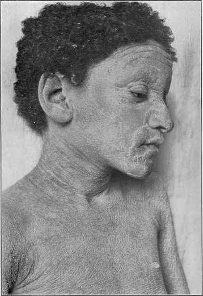
Pityriasis rubra pilaris in a mulatto girl, involving the entire surface. Began when
six or seven years old, and gradually extended, reaching generalization when ten years
old (at the time photograph was taken). Family free from skin disease, and brothers
and sisters, six in all, well and healthy. Under treatment improvement has slowly taken
place, so that a year ago, when last seen, then aged sixteen, not more than one-third of
the surface remained affected, the eruption then consisting of some large confluent scaly
areas and patches of closely crowded discrete follicular papules. The patient’s general
health has continued good throughout.
PITYRIASIS RUBRA PILARIS
233
or moderately thickened and scaly, but with no tendency to papular
formation. Ectropion of the lower eyelids sometimes ensues. The
lesions on the dorsal surfaces of the fingers usually remain discrete, and,
as likewise even in cases of considerable scaly development, are quite
distinctly pronounced.
The disease on both the scalp and the face is somewhat variable
as to degree, from a reddish, slightly scaly condition to that of some
thickening and marked scaliness, almost to the extent, on the latter
region, of producing a mask to the parts. The hairs of the scalp show
but little, if any, involvement. The nails, on the contrary, are often
brittle, rough, dull, striated, and they show a tendency to break and
crack. The disease is in most cases progressive, but after some months,
after reaching a variable extent, it may remain stationary for a time
and then advance again; or there may be periods of slight retrogression
and progression. It may be so extensive as to cover in most of or prac
tically the entire surface, in which event a generalized thickened and
inelastic condition of the skin is observed, covered with grayish scales,
usually moderate in quantity, and dry and harsh to the touch; the natural
lines of the skin are considerably exaggerated, and sometimes cracks about
the joints occur. In such instances the papular element is scarcely recog
nizable, although, as remarked, discrete papules are still, as a rule, to be
found on the backs of the phalanges. In less extensive cases large gray
ish plaques, irregularly shaped but commonly oblong, are to be seen on
different parts, especially the extremities, with outlying typical papules
and with some irregularly scattered over the general surface. In cases
of any extent the face, scalp, and hands rarely, if ever, escape. As a
rule, there are no subjective symptoms complained of; occasionally,
slight itching. The general health remains apparently unaffected.
Etiology and Pathology.—The disease is rare and the cause
is unknown. Neither heredity, sex, nor color seems to have any etio-
logic significance. It has been observed (Besnier, Hallopeau and
Brodier, Rasch) in quite young children, in Rasch‘s1 case at the age
of two and a half years; indeed, in the majority of cases it has its begin
ning in childhood or early youth. Of 5 cases under my observation,
1 was a mulatto girl of ten, in whom it developed when aged six; the
others were a woman aged twenty, a negress aged twenty-five, and two
men, aged respectively twenty-five and thirty. They were all apparently
in good health, and did not seem to be seriously inconvenienced, although
in 3 the disease was almost universal. There is an instance on record of
several cases in a family.2 As yet there is scant foundation for the theory
of its tuberculous causation.
The pathologic anatomy (Jacquet, Taylor, Ravogli, Hartzell,
Heidingsfeld, and others) discloses that the disease is a hyperkeratosis,
secondary inflammatory changes resulting. The essential and primary
hornification occurs in the epithelial lining of the orifice of the hair-
1 Rasch, Dermatolog. Centralblatt, No. 7, 1899, p. 199.
2 De Beurmann, Bith and Henyer, “Pityriasis rubra pilaire familial,” Annales,
1910, p. 609 (4 cases, two brothers and two sisters, in a family of six children, the
other two being free); the father was thought to have had it in a mild way, but this is
somewhat uncertain; two cousins were reported to have a similar condition.
234
INFLAMMATIONS
follicle, producing the papule; the projecting horny spine being due
to the collected mass of cornified epithelium within the follicle. All
the epidermic layers are markedly thickened, more especially the upper
corneous part.
The sweat-duct outlets may sometimes show similar, but relatively
insignificant, changes. Round-cell infiltration is noted about the hair-
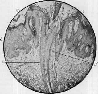
follicles and to a less extent in the papillae. In one of my cases sec
tions, from one of which the herewith illustration was taken, were
kindly made by Dr. Hartzell, who reported as follows:
“The epidermis was three or four times thicker than normal, the
increase in thickness being most marked in the corneous and prickle-

Fig. 45.—Pityriasis rubra pilaris: a, Thickened corneous layer; b, hypertrophied
rete; c, hair-follicle; d, cell infiltration about follicle; e, corneous plug in mouth of
follicle (section from the case herein pictured—section and photomicrograph by Dr. M.
B. Hartzell).
cell layers. The corneous layer, while everywhere thicker than normal,
was most markedly increased in and around the mouths of the hair-
follicles, in which it formed plugs of considerable size, projecting some
distance above the surface and extending well into the follicle. Many
of the cells of the rete mucosum showed greatly enlarged nuclei which
stained badly or, in many instances, not at all. The papillæ of the
corium were decidedly increased in length, were only slightly wider than
normal, and contained a moderate number of small round-cells with a
few ‘mastzellen.’ Along the entire length of the hair-follicles there was
a fairly abundant round-cell infiltration. Neither the sebaceous nor the
sweat-glands showed any appreciable alteration.”
Diagnosis.—In well-developed cases there is rarely difficulty in
PITYRIAS1S RUBRA FILARIS
235
the diagnosis; the follicular papules containing a horny projection and
broken hair-shaft, and usually to be found even in extensive cases, espe
cially on the dorsal aspects of the fingers, the thickened, harsh, rough,
and slightly or moderately scaly skin, the thickened palms and soles, the
marked scaliness of the face and scalp, constitute a picture usually quite
characteristic. In mild and moderate cases the papular lesions, gener
ally crowded together, with the features described, will be sufficiently
distinctive. When more or less general, it may show some similarity to
some cases of dermatitis exfoliativa, but in this latter there is rarely
material thickening, the skin is redder, and the scaliness more pro
nounced. It can scarcely be mistaken for psoriasis—in the latter
the beginning lesions, their character, and their growth by peripheral
extension, instead of by accretion of new papules as in pityriasis rubra
pilaris, and the absence usually of palmar and marked facial involvement,
are entirely different. The scaly plaques of lichen planus present some
features similar to the plaques of this malady, but the dark-red or vio
laceous tinge of the border and of outlying lesions in the former, and
the flattened, frequently umbilicated, characters of the discrete papules,
as a rule, always to be found, and the itchiness and almost invariable
absence of involvement of face, scalp, and palms, are distinguishing
characters.
Prognosis.—The disease is always persistent and rebellious to
treatment. Retrogression, however, may take place, and even com
plete recovery has been recorded; recurrence often ensues. The cases
under my care all improved, but not one was cured; only one was
under my care a sufficiently long time, however, to expect more than
betterment. In this last—the case shown in the illustration—the dis
ease, when the patient was seen several months ago, was still gradually
retrogressing.
Treatment.—The treatment of this malady consists essentially
in the administration of tonics, when necessary, sudorifics, and exter
nally bran, starch, or alkaline baths, and oils or ointment applications.
Exercise, proper food and living are, of course, of great value. Arsenic
is only exceptionally valuable in this disease; but, in view of Hebra's
experience as to its remarkable efficiency in continued large doses, it
might, in extensive cases, be tentatively tried in steadily increasing
dosage.1 Sodium cacodylate, in 1- to 3-grain (0.07-0.2) doses, adminis
tered hypodermically, proves sometimes of value. Thyroid extract
seemed of slight service in 1 case, but as the external treatment was
being carried out at the same time, it was doubtful to which the benefit
was due. Little2 had some effects from its use in one instance, begin
ning with 1½ grains (0.1) and increasing to 3 grains (0.2) three times
daily. Jaborandi or pilocarpin will sometimes have a favorable influence
by its action on the perspiratory glands. If the nutrition is poor, cod-
liver oil is a remedy of considerable value.
1 Heidingsfeld, Jour. Cutan. Dis., 1906, p. 371, found his 3 cases uninfluenced by
this drug in respect to its internal administration and the hypodermic injection of
sodium arsenate; but favorably influenced by hypodermic injections of atoxyl, and, to a
lesser extent, by cacodylic acid.
2 Little, Brit. Jour. Derm., 1900, p. 412.
236
INFLAMMATIONS
The external remedies are about the same as used in psoriasis and
in ichthyosis. In cases of decidedly irritable skin, bran or starch baths,
daily or three or four times weekly, prove serviceable. Alkaline baths
are most frequently to be used, and can be made up with the various
alkalis usually employed. Oil or ointment applications should be made
once or twice daily, as well as immediately after a bath. An oint
ment of salicylic acid, from 10 to 60 grains (0.654-4.) to the ounce
(32.), is one of the most efficient. Weak tar ointments are also at times
of service.1 Markedly thickened and hard areas can be treated by a
10 to 20 per cent, salicylic acid plaster, or this remedy can be applied
in collodion, 2 to 10 per cent, strength. Pyrogallol and resorcin salves
have been extolled, but the use of the former must be restricted to small
areas for fear of absorption. The scalp is to be shampooed frequently
with tincture of green soap, and an ointment or oil applied.
But first, if you want to come back to this web site again, just add it to your bookmarks or favorites now! Then you'll find it easy!
Also, please consider sharing our helpful website with your online friends.
BELOW ARE OUR OTHER HEALTH WEB SITES: |
Copyright © 2000-present Donald Urquhart. All Rights Reserved. All universal rights reserved. Designated trademarks and brands are the property of their respective owners. Use of this Web site constitutes acceptance of our legal disclaimer. | Contact Us | Privacy Policy | About Us |